


Транспозиция магистральных артерий
Содержание
Что такое D-транспозиция магистральных сосудов?
D-транспозиция магистральных сосудов - сложный врожденный порок сердца, характерным признаком которого является желудочково-артериальная дискордантность. Врожденная корригированная транспозиция - порок сердца, при котором наблюдается предсердно-желудочковая и желудочково -артериальная дискордантность.
Эмбриология
Данный порок относится к аномалиям конотрункуса. Конотрункус (bulbus, или bulbus cordis) – две тесно связанные эмбриональные структуры сердца – конус и артериальный трункус. Он является выносящим трактом сердца 2-3-недельного эмбриона. В норме на 4-5 неделе внутриутробной жизни плода конотрункус разделяется спиральной перегородкой на сравнительно равные по размеру крупные сосуды — аорту и легочный ствол.
Классическая теория развития этого порока предполагает нарушение формирования спиральной перегородки конотрункуса.
Альтернативная теория подразумевает, что в основе проблемы лежит недоразвитие подартериального конуса. В результате происходит фиброзное соединение между легочным и митральным клапанами (митрально-легочный контакт) – один из признаков транспозиции.
Анатомия
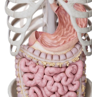
В нормальном сердце аорта отходит от левого желудочка, имеется митрально-аортальный фиброзный контакт и подлегочный конус. При классической ТМС легочное и системное кровообращение полностью разделены. Аорта, расположенная спереди, отходит от правого желудочка, а легочная артерия позади аорты — от левого желудочка.
Десатурированная кровь циркулирует в большом круге кровообращения, а насыщенная кислородом кровь — в малом.
Для выживания больного необходимо перемешивание крови, которое происходит на различных уровнях — через ДМПП, ДМЖП и ОАП. Часто данный порок сочетается с другими сердечными аномалиями. Другие варианты данного порока описаны в классификации.
Классификация
- D – ТМС с интактной межжелудочковой перегородкой - форма транспозиции, при которой аорта расположена кпереди и правее легочного ствола.
- D – ТМС с дефектом межжелудочковой перегородкой - форма транспозиции, при которой аорта расположена кпереди и правее легочного ствола, а также существует дефект межжелудочковой перегородки.
- Врожденная корригированная транспозиция, при которой перемещению подвергаются также морфологические левый и правый желудочки с соответствующими им атриовентрикулярными клапанами.
Гемодинамика
При D-ТМС кровоснабжение параллельное (в норме - последовательное). Кровь поступает по верхней и нижней полым венам в правые отделы сердца. Оттуда через аорту в системный круг кровообращения. И далее по замкнутому кругу обратно в правое предсердие.
По такой же схеме происходит кровоток в малом круге кровообращения: из легочных вен в легочную артерию. При отсутствии коммуникаций на уровне предсердий или желудочков - ребенок нежизнеспособен. При наличии сообщений на различных уровнях, кровь смешивается, уровень смешения зависит от возраста и размеров дефекта, чаще всего миксинг крови незначительный, что проявляется тяжелой гипоксемией.
Внутриутробно нарушение кровообращения характеризуется сниженным насыщением кислородом крови, поступающей в головной мозг и восходящую аорту. При тяжелой обструкции выводного отдела правого (системного) желудочка, кровоток в большом круге является дуктус-зависимым. Закрытие ОАП сопровождается быстро прогрессирующим ухудшением системной перфузии. При корригированной ТМС гемодинамика не нарушается, однако со временем функция правого желудочка начинает ухудшаться из-за его постоянной работы на системный круг кровообращения.
Диагностика
- ЭхоКГ, КТ, МРТ. Визуализация дефекта и получение функциональных данных, необходимых для проведения лечения.
- ЭКГ. Электрическая ось сердца отклонена вправо. Гипертрофия правого желудочка при простой ТМА. Гипертрофия обоих желудочков регистрируется при сопутствующих ДМЖП, ОАП.
- Рентгенография органов грудной клетки. Характерны кардиомегалия, яйцеобразная форма сердечной тени с узким верхним средостением (магистральные сосуды расположены в переднезаднем направлении). Легочной рисунок усилен.
- Катетеризация сердца. Для выполнения баллонной атриосептостомии, также используют с диагностической целью для измерения давления в левом желудочке, уточнения деталей анатомии порока и особенностей отхождения коронарных артерий.
Клинические проявления
Для больных с ТМС характерно сочетание цианоза с застойной сердечной недостаточностью, одышкой, трудностями кормления в период новорожденности.
Аускультативно: II тон громкий, нерасщепленный. Шумы отсутствуют при интактной межжелудочковой перегородке. Систолический шум слышен у больных с сопутствующим ДМЖП. Иногда выслушивается мягкий систолический шум выброса при стенозе легочной артерии. При застойной сердечной недостаточности увеличена печень.
При исследовании газов крови отмечается тяжелая артериальная гипоксемия, которая не устраняется ингаляцией кислорода.
Лечение
Оперативному вмешательству обычно предшествуют баллонная атриосептостомия (операция Rashkind). Пациентам у которых отсутствует сообщение на уровне предсердий или желудочков (ДМПП,ДМЖП) баллонная атриосептостомия должна быть выполнена сразу после их поступления в кардиохирургический центр.
На предоперационном этапе необходимо провести коррекцию метаболического ацидоза, гипогликемии и гипокальциемии, начать инфузию простагландина для улучшения насыщения артериальной крови кислородом путем поддержания проходимости ОАП. На данный момент существует мнение об отсутствии необходимости в инфузии простагландина при наличии функционирующего ДМПП. При наличии у пациента достаточного по размеру ДМПП, коррекция выполняется без предшествующих вмешательств. Выбор оперативного вмешательства зависит от наличия или отсутствия сопутствующих пороков сердца, анатомии коронарных артерий, типа транспозиции (D/L - ТМС).
На предсердном уровне осуществляют перераспределение потоков крови, используя один из двух вариантов — операцию Senning (легочный и системный венозный возврат перенаправляют с использованием тканей предсердий) или операцию Mustard (с помощью перикардиальной заплаты).
На данный момент эти операции применяются для лечения корригированной транспозиции магистральных сосудов (как часть операции Double Switch). К анатомической коррекции порока относят операцию артериального переключения.
Операцию артериального переключения (Jatene) выполняют в неонатальном периоде (3-5 сутки), пока левый желудочек еще не утратил способности осуществлять системное кровообращение, как во внутриутробном периоде. Суть операции заключается в перемещении аорты вместе с коронарными артериями на место легочного ствола, а ствола, соответственно, на место аорты. ТМС с ДМЖП и выраженным субаортальным стенозом корригируют с помощью операции Damus-Kaye-Stansel (соединяют проксимальную часть легочного ствола с восходящей аортой, закрывают ДМЖП и соединяют правый желудочек с дистальной частью легочной артерии с помощью синтетических или биологических протезов).