


Left ventricle outflow tract obstruction
Table of contents
What is Left Ventricular Outflow Tract Obstruction?
Left ventricular outflow tract obstruction may occur because of aortic valve stenosis, subaortic stenosis, and supravalvular stenosis.
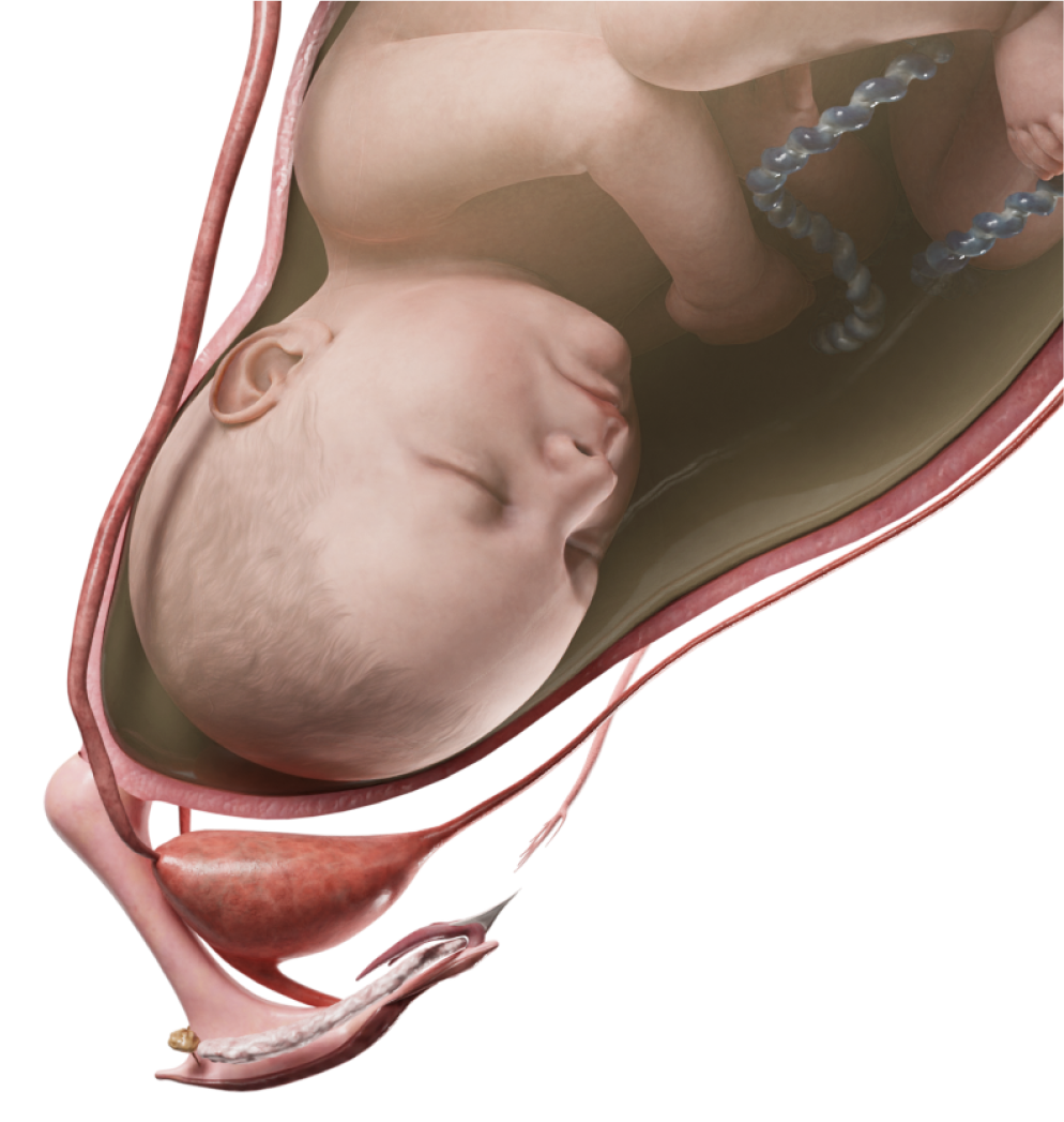
Embryology
At the 4th week of embryogenesis, the common arterial trunk is divided into two main vessels - aorta and pulmonary artery - by the formation of an internal septum due to endocardial invagination along two grooves. Separation occurs between the IV and VI aortic arches and continues down. A continuation of the aortic canal are the vessels originating from the IV aortic arches (the final aortic arch and the brachiocephalic trunk are formed from it), and a continuation of the pulmonary canal are the vessels originating from the VI aortic arches (the open arterial duct and part of the right pulmonary artery are formed from it).
The right ventricle connects to the VI aortic arches (pulmonary arteries), and the left ventricle connects to the III and IV arches. At a later stage of embryo development, on the side of the inner wall of the great vessels beginning thickenings, consisting of mesenchymal cushions, are formed. from which four valves of arterial valves are formed. Four leaflets of arterial valves are formed from these thickenings. After the bulbus is divided into two excretory tracts, one pair of leaflets is further divided into two parts. As a result, six valve leaflets are formed, of which three leaflets form the pulmonary valve, and the other three - the aortic valve. At the same time, AV valves are formed due to thickening of the mesenchyme (the so-called endocardial cushions).
Numerous studies have shown a familial predisposition to aortic valve defects.
Violations of cardiogenesis at the stage of formation of the outflow tract of the left ventricle can lead to the defects of this group.
Classification
- Aortic valve stenosis;
- Subaortic stenosis;
- Discrete subaortic stenosis;
- Tunnel subaortic stenosis;
- Hypertrophic obstructive cardiomyopathy;
- Structural subaortic stenosis;
- Supravalvular aortic stenosis.
Aortic valve stenosis
Anatomy
Valve stenosis is represented by a bicuspid or tricuspid valve. The valve cusps are fused along the commissures, with a small hole in the center. In addition, the sups are often thickened, incompetent.
Hemodynamics
Narrowing the valve creates a pressure gradient between the left ventricle and the aorta during systole.
As the blood flow through a narrowed valve is turbulent, the gradient is not a simple linear function of the flow. It is directly proportional to the cross-sectional area of the valve, so doubling the volume of blood flow through the valve increases the gradient by four times.
The magnitude of the gradient depends on the contractility of the left ventricle and the resistance of the peripheral vessels. To assess the hemodynamic severity of obstruction, it is necessary to measure the volume of blood flow through the valve and at the same time the gradient.
The average pressure in the left atrium is usually normal. End-diastolic pressure in the left ventricle is at the level of the upper normal limit. Its increase indicates a deterioration in the function of the left ventricle or pronounced hypertrophy and a decrease in the compliance of this chamber.
Myocardial blood supply is reduced, despite patency of the coronary arteries.
Workup
- Echocardiography, CT. Visualization of the defect.
- Chest X-ray. Expansion of the shadow of the aorta can be found.
- ECG. Characteristic changes are absent or there are signs of left ventricular hypertrophy.
- Cardiac catheterization and angiography. It is used to clarify the localization and determine the degree of aortic stenosis.
Clinical presentation
Unlike other heart defects, severe aortic stenosis can occur for a long time without clinical manifestations. Children grow and develop normally.
Patients may complain of fatigue, shortness of breath on exertion, chest pain and dizziness. Less often, they complain of abdominal pain, profuse sweating, and nosebleeds. Fatigue and dyspnea during physical activity indicate a moderate stenosis.
Dizziness during physical activity is a sign of severe aortic stenosis. It is explained by the inability of the left ventricle to increase blood flow in order to maintain adequate cerebral blood flow. Anginal pain occurs due to the inconsistencies in oxygen delivery with myocardial needs.
If aortic stenosis is manifested by severe symptoms during the neonatal period, the definition of “critical” is associated with an immediate threat to life and the need for emergency intervention. In most cases, there is a bicuspid aortic valve.
Auscultation. An aortic output sound is heard at the apex of the heart if the valve is movable. The opening cusps generate the sound; its intensity does not change during breathing. The presence of an IV cardiac tone indicates severe obstruction, because hypertrophy prevents the passive filling of the left ventricle. A loud, rough systolic murmur of aortic stenosis is also heard, better heard at the base of the heart.
Treatment
A deformed aortic valve is a potential site of bacterial infection, therefore, constant attention must be paid to the prevention of infection, regardless of the severity of the narrowing.
Methods of surgical treatment are adapted to the condition of patients, age and form of the defect.
- The method of choice for infants in critical condition is balloon valvuloplasty.
- Open-heart aortic valvotomy is used for older children.
- Aortic valve replacement may be unavoidable in the presence of a unicuspid valve or dysplastic bicuspid valve, after a previous valvotomy, due to progressive degenerative changes and calcification.
- Pulmonary valve autotransplantation into the aortic position and restoration of the right ventricular outflow tract with a homograft (the Ross operation).
- Patients with severe stenosis of the valve annulus or tunnel-like obstruction need an aortic root extension (the Konno operation) in combination with valve prosthetics or Ross surgery (the Ross – Konno operation).
Subaortic stenosis
Anatomy
Subaortic stenosis is a subvalvular narrowing of the left ventricular outflow tract, most often in the form of a fibrous membrane or ring. There are several anatomical forms of the defect: a discrete membrane, structural subaortic stenosis and a tunnel form. Discrete subaortic membrane - narrowing of the LVOT component due to a fibrous membrane with a centrally located opening. Structural subaortic stenosis is a circular fibrous thickening in the form of ridges. In case of the tunnel form, a fibromuscular fold encircles LVOT in form of a collar. Obstructive HCM also belongs to the group of defects with subaortic stenosis.
Hemodynamics
The obstacle in the systemic outflow determines hemodynamic disorders. The gradient of systolic pressure in this defect occurs inside the ventricle itself and the main compensation mechanism is a pressure increase in the left ventricle. Subsequently, myocardial hypertrophy develops, left atrium pressure increases, which ultimately leads to left ventricular failure.
Workup
- Echocardiography, CT. Visualization of the defect.
- Chest X-ray. There is an expansion of the left ventricle. The heart has an aortic configuration with a pronounced "waist".
- ECG. Signs of left ventricular hypertrophy.
Clinical presentation
Isolated subaortic stenosis is rare among newborns and young children.
In most cases, it occurs as a concomitant pathology and signs of the disease are “masked” by a major defect in early age. In older children (after 3 years) and in some adults, even severe stenosis can be asymptomatic. The main complaints of patients are dyspnea, fatigue and heart pain. One of the clinical features of the defect is syncope.
Loud ascending heartbeat on the apex, discrepancy between it and a rare small pulse. A loud systolic murmur above the base of the heart or in the third intercostal space on the left with irradiation to the neck and jugular fossa is heard. Systolic tremor above the carotid arteries is characteristic.
Treatment
Surgery is the only effective treatment for this defect. The Konno operation and its modifications, resection of subvalvular stenosis of the aortic valve are used. Endovascular techniques are also often used: transluminal balloon dilation.
Supravalvular aortic stenosis
Anatomy
Supravalvular aortic stenosis is a local or diffuse stenosis of the ascending aorta at and above the sinotubular area. The morphological basis of the stenosing segment of the aorta is endothelial fibrosis, extreme proliferation and disorganization of the mesothelium. Supravalvular aortic stenosis is divided into three anatomical subtypes: the form of a fibrous diaphragm, local hourglass stenosis or diffuse aortic hypoplasia.
Hemodynamics
As with other types of the left ventricle outflow tract obstruction, hypertrophy and overload of the left ventricle, as well as the presence of a high-pressure gradient between the aorta and the left ventricle characterize the pathological process. Unlike subvalvular and valvular aortic stenosis, the coronary arteries are located proximal to the site of stenosis and are under the influence of high pressure in the ventricle.
Therefore, coronary vessels are often dilated, convoluted, with signs of early arteriosclerosis.
Workup
- Echocardiography, CT. Visualization of the defect.
- ECG. Signs of left ventricular hypertrophy.
Clinical presentation
Signs of supravalvular stenosis are similar to those of other types of aortic stenosis, although anginal pain is more common.
Despite the fact that auscultatory data are similar for all types of aortic stenosis, there is no systolic click above the aortic valve and the epicenter of systolic murmur is higher than with subaortic stenosis. The sound of aortic valve closure is accentuated due to increased pressure in the aorta proximal to the site of stenosis. Blood pressure on the right hand may be higher than on the left, which may be due to narrowing of the aortic arch or subclavian artery. The pulse on the left hand can also be weakened.
Treatment
As the child grows, progression of aortic changes occurs, a characteristic clinical picture of the defect develops, and there is a need for its surgical correction, usually aged 2 to 20-30 years. Indications for surgical treatment are the presence of clinical symptoms, the pressure gradient in the area of stenosis of more than 50 mm Hg and impaired coronary circulation. For surgical correction, methods of plastic expansion of the ascending aorta are used.