


Аорто-желудочковый туннель
Содержание
Что такое Аорто-левожелудочковый или аорто-правожелудочковый туннель?
Аорто-левожелудочковый или аорто-правожелудочковый туннель - чрезвычайно редкий ВПС, при котором имеется прямое сообщение между левым или правым желудочком и восходящей частью аорты в обход аортальных клапанов.
Эмбриология
Считается, что образование аномалии связано с врожденным истончением передней стенки выводного тракта левого желудочка в месте, где правый аортальный синус соприкасается с мембранозной перегородкой.
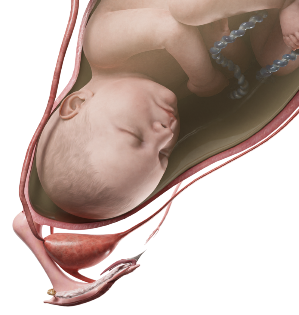
Данный порок можно обнаружить с 18-й недели беременности; однако в этих условиях это трудно диагностировать. Поэтому чаще всего этот дефект можно заподозрить между 22-й и 24-й неделями беременности.
Анатомия
Чаще встречается аорто-левожелудочковый туннель. Восходящая часть аорты резко расширена.
От ее передне-левой или передне-правой поверхности над уровнем коронарных отверстий артерий отходит сосудистое образование, покрытое эпикардом. Канал может иметь форму трубки или аневризмы. Аортальное отверстие обычно расположено на уровне комиссуры клапана и отделено от правого синуса Вальсальвы выступающим поперечным гребнем. Туннель расположен кнаружи от фиброзного кольца, справа от легочного ствола, где теряется в толще миокарда вблизи межжелудочковой перегородки.
Туннель может оттеснить вправо выводную часть межжелудочковой перегородки, создавая подлегочный стеноз. Изредка туннель может открываться в инфундибулярный отдел правого желудочка.
На разных уровнях по ходу туннеля стенка его может быть аневризматически расширенной. Порок может сопровождаться ДМЖП, аортальным стенозом или атрезией аорты с гипоплазией левого желудочка и легочным клапанным и подклапанным стенозом.
Классификация
- Из правого синуса Вальсальвы;
- Из левого синуса Вальсальвы.
Гемодинамика
Гемодинамические нарушения связаны с регургитацией крови из аорты в желудочек. Изменения гемодинамики зависят также от сочетания данного порока с другими врожденными пороками сердца.
Туннель не создает препятствия для свободного тока крови во все фазы сердечного цикла. Определенная часть крови во время систолы поступает назад в желудочек. Соответственно желудочек испытывает объемную перегрузку. У данных пациентов быстро развиваются кардиомегалия и сердечная недостаточность.
При отсутствии каких-либо сопутствующих пороков, гемодинамические нарушения связаны с постоянной недостаточностью выхода крови из аорты при сохранности имеющихся аортальных створок.
Диагностика
- ЭКГ. выраженная в различной степени гипертрофия левого желудочка и левого предсердия.
- Допплер – ЭХОКГ, КТ. Обнаружение туннеля (визуализация дефекта) и проведение дифференциальной диагностики.
- Рентгенография органов грудной клетки. признаки застойной сердечной недостаточности, увеличение размеров сердца, расширение восходящей аорты, в некоторых случаях выбухание правого аортального синуса.
- Катетеризация сердца с аортографией. для исключения сопутствующих внутрисердечных аномалий.
- ЭхоКГ, допплер-кардиография и аортография позволяют отличить этот порок:
- от недостаточности аортального клапана по отсутствию ретроградного тока через аортальный клапан;
- от коронарного свища, открывающегося в левый желудочек, — по наличию нормальных правой и левой главных коронарных артерий;
- от сопутствующего ДМЖП — по отсутствию шунта крови через дефект;
- от разрыва синуса Вальсальвы — по переднему расположению туннеля и отсутствию дилатации синусов аорты.
Клинические проявления
Так как порок создает нагрузку на сердце уже во внутриутробном периоде, выраженная сердечная недостаточность может возникнуть вскоре после рождения ребенка. Проявления объемной перегрузки сердца: тахикардия, одышка, увеличение печени.
В целом клиническая картина напоминает недостаточность аортального клапана. Выслушиваются систоло-диастолические шумы слева от грудины и на верхушке сердца. Пульс высокий, диастолическое артериальное давление снижено.
Лечение
Диагноз аорто-желудочкового туннеля является показанием для операции, при этом она должна быть выполнена как можно раньше.
Хирургические подходы предполагают закрытие аортального и\или желудочкового конца туннеля. Закрытие обоих концов предпочтительнее, так как предотвращает дальнейшую дилатацию туннеля. Особое внимание уделяют коронарным артериям и аортальному клапану для исключения их повреждений.
В отдаленном послеоперационном периоде у части пациентов имеется деформация аортального клапана, и в последующем требуется его пластика или протезирование. Вовремя проведенные операции приводят к быстрому восстановлению функции сердца и отсутствию проблем, связанных с аортальным клапаном.